VIDEO: Balance Assessment of Child with Hemifacial Microsomia (HFM) and Delayed Neuromotor Function
Published on: June 18, 2014
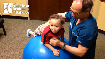
Case Study: a 3 year 8 month old male referred for balance assessment by a pediatric ENT. The child has a right-sided hemifacial microsomia (HFM). It has been determined from CT scan that inner ear anatomical structure is intact. Pre, peri and post birth history is unremarkable. He is the middle child (siblings are 6 and 2 years of age). He appeared to have normal maturational motor milestones until age 6-9 months when age-appropriate development seemed to lapse. The parents report that there has been no additional degradation since about age two years, where he seems to have plateaued. Presently, he can only crawl, not walk and he is not toilet trained. There is poor muscle development of the lower extremities and he cannot stand. There is significant speech and language delay, despite normal hearing acuity determined with objective testing e.g. BAER and DPOAE. He does vocalize, but only guttural sounds.
The ideal objective vestibular test would be c-VEMP, which could not be conducted due to the right microsomia and a concurrent left middle ear effusion (conductive hearing loss). Therefore, a behavioral motor-milestone assessment was conducted, as seen in the video, until such time as the left ear effusion is resolved and the c-VEMP may be performed. It is not anticipated however, based on the normal behavioral findings that c-VEMP would be abnormal or provide additional diagnostic data which would change the triage or management strategies. The child is presently receiving physical, occupational and speech therapy. The results of the behavioral balance assessment did not reveal any indicators that there is a likely labyrinthine contribution to his poor motor control function.
There are no clear reports in the literature, which are linked to balance dysfunction in the FHM population. The nature of the degenerative decline and condition suggests a likely complex syndromic co-morbidity, which has not yet been identified.
References: condition summary on craniofacial microsomia.
-
- Gene Review: Craniofacial Microsomia
-
- Gougoutas AJ, Singh DJ, Low DW, Bartlett SP. Hemifacial microsomia: clinical features and pictographic representations of the OMENS classification system. Plast Reconstr Surg. 2007 Dec;120(7):112e-120e. Review.
-
- Hartsfield JK. Review of the etiologic heterogeneity of the oculo-auriculo-vertebral spectrum (Hemifacial Microsomia). Orthod Craniofac Res. 2007 Aug;10(3):121-8. Review.
-
- Johnson JM, Moonis G, Green GE, Carmody R, Burbank HN. Syndromes of the first and second branchial arches, part 1: embryology and characteristic defects. AJNR Am J Neuroradiol. 2011 Jan;32(1):14-9. doi: 10.3174/ajnr.A2072. Epub 2010 Mar 18. Review.
-
- Johnson JM, Moonis G, Green GE, Carmody R, Burbank HN. Syndromes of the first and second branchial arches, part 2: syndromes. AJNR Am J Neuroradiol. 2011 Feb;32(2):230-7. doi: 10.3174/ajnr.A2073. Epub 2010 Apr 1. Review.
-
- Keogh IJ, Troulis MJ, Monroy AA, Eavey RD, Kaban LB. Isolated microtia as a marker for unsuspected hemifacial microsomia. Arch Otolaryngol Head Neck Surg. 2007 Oct;133(10):997-1001.
-
- Rahbar R, Robson CD, Mulliken JB, Schwartz L, Dicanzio J, Kenna MA, McGill TJ, Healy GB. Craniofacial, temporal bone, and audiologic abnormalities in the spectrum of hemifacial microsomia. Arch Otolaryngol Head Neck Surg. 2001 Mar;127(3):265-71.
-
- Tasse C, Böhringer S, Fischer S, Lüdecke HJ, Albrecht B, Horn D, Janecke A, Kling R, König R, Lorenz B, Majewski F, Maeyens E, Meinecke P, Mitulla B, Mohr C, Preischl M, Umstadt H, Kohlhase J, Gillessen-Kaesbach G, Wieczorek D. Oculo-auriculo-vertebral spectrum (OAVS): clinical evaluation and severity scoring of 53 patients and proposal for a new classification. Eur J Med Genet. 2005 Oct-Dec;48(4):397-411. Epub 2005 Jun 8.
-
- Vendramini-Pittoli S, Kokitsu-Nakata NM. Oculoauriculovertebral spectrum: report of nine familial cases with evidence of autosomal dominant inheritance and review of the literature. Clin Dysmorphol. 2009 Apr;18(2):67-77. doi: 10.1097/MCD.0b013e328323a7dd. Review.
-
- Werler MM, Starr JR, Cloonan YK, Speltz ML. Hemifacial microsomia: from gestation to childhood. J Craniofac Surg. 2009 Mar;20 Suppl 1:664-9. doi: 10.1097/SCS.0b013e318193d5d5. Erratum in: J Craniofac Surg. 2009 Sep;20(5):1629-30.